(Dr. Deepak Chhabra, Dr. K Mrityunjay)
Introduction
In response to the demand for a clinical definition of fibromyalgia syndrome (FMS), the Expert Consensus Panel, selected by Health Canada, established clinical criteria that encompass the potential pathophysiological dysfunctions, and developed an integrative approach to the diagnosis and treatment of FMS.
Classification
The prominent feature of FMS is chronic, widespread musculoskeletal pain, but it is usually accompanied by numerous other multisystemic dysfunctions. Fibro refers to the fibrous tissue, myo refers to the muscles and algia refers to pain. Fibromyalgia is assigned number M79.0 and is classified as nonarticular rheumatism in the World Health Organization’s International Classification of Diseases (ICD). FMS is in the “generalized” category of the large group of softtissue pain syndromes, implying that a systemic process involves the musculoskeletal system globally. Compelling evidence of physiological and biochemical abnormalities identifies FMS as a distinct pathophysiological clinical disorder.
Etiology
Before the onset of FMS, most patients enjoyed an active, healthy lifestyle. There is consistent documentation that a physical trauma, particularly a whiplash or spinal injury, can trigger FMS in some patients. Other associated physical traumas include surgery, repetitive strain, childbirth, viral infections and chemical exposures. A genetic predisposition may be suggested in cases where more than one separated family member is afflicted. Some cases of FMS have a gradual onset with no obvious cause.
Prevalence
Canadian epidemiological studies indicate between 2 and 10 percent of the general population, or between 600,000 and 3 million people, have FMS. It is two to five times more prevalent than rheumatoid arthritis. It affects all age groups, including children, all racial/ethnic groups, and all socioeconomic strata. There is a higher prevalence in females. The generally more flexible, delicate skeletons, less massive muscles, and narrower spinal canals of females may make them more prone to neck and spinal injuries. A whiplash injury study suggests those with persistent symptoms have a significantly narrower cervical spinal canal (particularly females) 9. Females produce more neurotransmitters that increase pain signals and fewer neurotransmitters that decrease pain signals than males. Both the direction and magnitude of the brain’s response to pain differs in males and females, with females being more sensitive to pain.
Natural Course
An eight year multicentre study suggests that generally once FMS has been established, patients do not improve symptomatically and there is a slight worsening of functional disability 7. A 15 year study8 indicates that all patients in the study still have FMS but there is some variation in symptom severity. Individual prognosis must remain a clinical estimate because the prognosis for an individual patient cannot be predicted accurately with certainty.
Diagnostic Criteria
The Expert Consensus Panel adopted the 1990 American College of Rheumatology criteria, which have good sensitivity and specificity, and also included a broader spectrum of the potential symptomatic expressions of FMS to form a clinical working case definition.
| Diagnostic Criteria for Fibromyalgia | |
| The two compulsory pain criteria (adopted from the American College of Rheumatology 1990 Criteria) are merged with Additional Clinical Symptoms and Signs to expand the classification of FMS into a Clinical Working Case Definition of FMS. | |
| 1. Compulsory HISTORY of widespread pain. Pain is considered widespread when all of thefollowing are present for at least three months:- Pain in both sides of the body- Pain above and below the waist (including low back pain)- Axial skeletal pain (cervical spine, anterior chest, thoracic spine or low back). Shoulder and buttock involvement counts for either side of the body. “Low back” is lower segment. | |
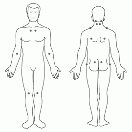
| 2. Compulsory PAIN ON PALPATION at 11 or more of the following 18 tender point sites:- Occiput (2): at the suboccipital muscle insertions.- Low cervical (2): at the anterior aspects of the intertransverse spaces (the spaces between the transverse processes) at C5 – C7.- Trapezius (2): at the midpoint of the upper border- Supraspinatus (2): at origins, above the scapular spine near its medial border.- Second rib (2): just lateral to the second costochondral junctions, on the upper rib surfaces.
– Lateral epicondyle (2): 2 cm distal to the epicondyles (in the brachioradialis muscle). – Gluteal (2): in upper outer quadrants of buttocks in the anterior fold of muscle. – Greater trochanter (2): posterior to the trochanteric Prominence. – Knee (2): at medial fat pad proximal to the joint line.
|
|
| 3. Additional Clinical Symptoms and Signs: In addition to the compulsory pain and tendernessrequired for research classification of FMS, many additional clinical symptoms and signs can contribute importantly to the patients’ burden of illness. Two or more of these symptoms are present in most FMS patients by the time they seek medical attention. On the other hand, it is uncommon for any individual FMS patient to have all of the associated symptoms or signs. As aresult, the clinical presentation of FMS may vary somewhat, and the patterns of involvement may eventually lead to the recognition of FMS clinical subgroups. These additional clinical symptoms and signs are not required for research classification of FMS but they are still clinically important. For these reasons, the following clinical symptoms and signs are itemized and described in an attempt to expand the compulsory pain criteria into a Clinical Case Definition of FMS:- Neurological manifestations: Neurological difficulties are often present such as hypertonicand hypotonic muscles; musculoskeletal asymmetry and dysfunction involving muscles, ligaments and joints; atypical patterns of numbness and tingling; abnormal muscle twitch response, muscle cramps, muscle weakness, and fasciculations. Headaches, temporomandibular joint disorder, generalized weakness, perceptual disturbances, spatial instability, and sensory overload phenomena often occur.
– Neurocognitive manifestations: Neurocognitive difficulties usually are present. These include impaired concentration and shortterm memory consolidation, impaired speed of performance, inability to multitask, easy distractibility, and/or cognitive overload.
– Fatigue: There is persistent and reactive fatigue accompanied by reduced physical and mental stamina, which often interferes with a patient’s ability to exercise.
– Sleep disturbance: Most FMS patients experience nonrefreshing sleep. This is usually accompanied by sleep disturbances including insomnia, frequent nocturnal awakenings, nocturnal myoclonus, and/or restless leg syndrome.
– Autonomic and/or neuroendocrine manifestations: These manifestations include cardiac arrhythmias, neurally medicated hypotension, vertigo, vasomotor instability, sicca syndrome, temperature instability, hot/cold intolerance, respiratory disturbances, intestinal and bladder motility disturbances with or without irritable bowel or bladder dysfunction, dysmenorrhea, loss of adaptability and tolerance for stress, emotional flattening, lability, and/or reactive depression.
– Stiffness: Generalized or even regional stiffness that is most severe upon awakening and typically lasts for hours usually occurs, as in active rheumatoid arthritis. Stiffness can return during periods of inactivity during the day.
|
|
Application Notes
- Digital palpation examination is performed with an approximate force of 4 kg/1.4 cm2 (standardize on a weight scale), which will partially blanch the blood from under the thumbnail. The patient must state that the palpation was painful to be considered “positive”. “Tender” is not considered “painful”.
- Validity of the two compulsory pain criteria for the purposes of research study yielded 88.4% sensitivity and 81.1% specificity.
- Focus of the clinical definition: The following hourglass diagram indicates the steps in first narrowing the compulsory criteria to establish FMS and then expanding the spectrum of symptoms and signs and the distress they can cause to establish the total illness burden.

General Considerations in Applying the Clinical Case Definition
- Determine the patient’s total illness burden by assessing all of the patient’s symptoms and their impact on the patient’s lifestyle demands, occupations, etc.
- Coherence of symptoms: Symptoms should fit a pattern that is identifiable as FMS.
- Identify secondary symptoms and aggravators. Symptom dynamics and interactions and the effects of aggravators should be noted.
- Quantify the severity of major symptoms, and their impact on lifestyle. When the symptom severity and severity hierarchy profile chart is completed every six months or so, it will help orient treatment, assess its effectiveness, and assist in assessing prognosis and disability. Impact on lifestyle should be compared to the patient’s premorbid health and activity level.
Signs & Symptoms
- Pain and Neurological Manifestations
A comprehensive biological model suggesting dysregulation among the central nervous system (CNS), autonomic nervous system (ANS), and body organs systems is emerging. Functional Imaging studies support the theory that many signs and symptoms of FMS originate from dysfunction of the CNS and altered processing of sensory input. Chronic generalized pain may be primarily a central nervous system phenomenon, an abnormality in the brain’s sensory perception and processing of pain, even though the onset may be related to a peripheral event. Neurochemical factors may play an important role in the amplification and distortion of pain signals in the nociceptive process.
The pain of FMS may be described as burning, searing, sharp, shooting, stabbing, throbbing, deep aching, tingling, feeling bruised all over, aching in bones, exhausting, etc. or any combination of these. Pain and fatigue may be induced by exercise and there is a slow recovery period. Myofascial trigger points are commonly found in FMS patients and myofascial pain syndrome (MPS) should be considered a concomitant diagnosis.
a. Characteristics of FMS Pain
- Allodynia is a reduced pain threshold from a stimulus which would not normally be painful.
- Hyperalgesia is an abnormally high sensitivity and perceived greater intensity of pain to a stimulus that would be expected to produce some pain.
- Persistent pain: The duration of pain from a stimulus is longer than would be expected.
- Pronounced summation effect and afterreaction to a painful stimulus often occurs.
- Hyperalgesia in skin: Affected dermatomes produce more pain when a pin is drawn across the skin.
- Tenderness: Pain that does not radiate to a distant site may be experienced on palpation of tender points and is independent of widespread pain. Tender points are generally where ligaments, tendons, and muscles attach to the bone.
Muscles, ligaments, tendons, fascia, and the periosteum are sensitive to pain. Injuries to ligaments, such as a whiplash injury, can overstretch and fray their cable like structure. Ligaments are difficult to heal because they have a poor blood supply, particularly where they attach to the bone. Lax ligaments allow the joint to move beyond its normal range of motion, which compresses or irritates sensory nerves, and causes pain, numbness and/or tingling. The muscles around the joint tend to react to these pain signals by contracting and becoming chronically taut in their attempts to stabilize the joint and prevent further damage.
b. Other Features of FMS Pain:
- Widespread pain: Pain that is felt bilaterally, as well as above and below the waist, is considered widespread. A soft tissue injury, such as a whiplash injury, may initiate local or regional pain that over the course of months becomes generalized, widespread pain with positive tender points. This suggests that FMS involves abnormalities in the pain processing interaction between the peripheral and CNS.
- Nonanatomical distribution: Global or regional nonanatomical pain may occur unexpectedly, fluctuate, and is often migratory.
- Delay in onset after prodromal injury or event
- Diffuse arthralgia: The pain in joints occurs without redness and swelling of the joints, which differentiates it from rheumatoid arthritis (RA).
- Shortness of breath, and atypical chest pain resembling angina
- Low back pain may be accompanied by shooting, sciaticalike leg pain. Concomitant piriformis pain and compression of the sciatic nerve may occur.
- Leg cramps occur in approximately 40% of patients.
- Generalized stiffness: Studies suggest that morning stiffness of more than 15 minutes duration occurs in 79% to 83% of patients. Stiffness can reoccur during the day, usually after periods on nonactivity.
- Chronic headache: Approximately 50-60% of patients experience severe tension headaches involving cervical and shoulder girdle muscle contraction. Migraine like headaches may occur and may be preceded by visual disturbances.
- Temporomandibular joint disorder is common and is usually caused by chronic contraction of the muscles involved in the joint movement in FMS patients.
c. Other neurological manifestations
Mismanagement of sensory information may be due to dysfunction of neurotransmitters/ receptors and abnormal gating (the process whereby the prefrontal cortex assigns relative importance to sensory input) resulting in dysregulation of the signal to noise ratio. This dysregulation may result in a lower tolerance of noxious stimuli.
- Hypersensitivity to vibration
- Positive Romberg test
- Abnormal tandem gait and interference aggravation. Even when tandem gait and serial 7 subtraction test results are normal when done independently, many patients have difficulty or are unable to do them concurrently.
- Abnormal twitch response associated with myofascial pain syndrome.
- Muscle and/or generalized weakness and fasciculations
- Dysesthesia: Atypical patterns of numbness (approximately 65% 24 ) and tingling often occur in the feet and hands and may be accompanied by swelling. Patients may undergo unsuccessful surgery for carpal tunnel syndrome. Therefore, such surgery should not be done unless there is confirmed median nerve injury and thenar wasting or weakness of opponens strength.
- Perceptual disturbances, and temporal and spatial instability: Difficulty with visual accommodation and focusing, loss of depth perception, and an inability to distinguish figure/ground may result in the patient appearing clumsy and an inability to accommodate walking on uneven surfaces. Temporal instability may result in difficulty in sequencing actions.
- Overload phenomena: Patients may be hypersensitive to noise, light, odours, speed and to mixed sensory modalities. Cognitive, motor, perceptual, and emotional overload causes a worsening of other symptoms and may result in patients becoming temporarily immobilized.
- Cervical cord compression myelopathy may produce local dysfunction at the implicated cervical roots and abnormal long tact signs. A thorough neurological investigation including MRI of the foramen magnum and cervical spine should be done on patients with abnormal neurological presentations. Early diagnosis and treatment of spinal stenosis give better results.
- Neurocognitive Dysfunction
Slowed processing of information may be related to sleep dysfunction, chronic pain, headaches and cognitive fatigue. A dysfunction of the prefrontal cortex, which helps regulate the hippocampus in its production of new memories, may result in a failure to integrate information, or misinterpret information as being novel because the cognitive context is absent or unavailable Dysfunction of REM sleep and hippocampal neural firing during slow wave sleep may play a role in difficulties in concentration and attention, and ease of distraction resulting in poor initial learning and memory consolidation. Concentration difficulties may also be related to hypoactivity of the frontal lobes when awake. Patients who also meet the criteria of myalgic encephalomyelitis/chronic fatigue syndrome (ME/CFS) generally have more severe neurocognitive problems. Symptoms vary but often reflect slowed cognitive functioning related to cognitive fatigue. “Fibro fog” is a term often used to refer to the confusion, forgetfulness, and difficulties with concentration, word retrieval and speaking, short term memory consolidation, and susceptibility to interference that FMS patients often experience. Physical and cognitive overload and/or fatigue may lead to a worsening of other symptoms.
- Fatigue
Patients generally awaken feeling more exhausted than when they went to bed. Postexertional fatigue, weakness, increased pain and stiffness, and worsening of other symptoms are typical. Onset may be immediate or delayed and recovery time is abnormally long. Fatigue may also appear unexpectedly or inappropriately, and may be migratory in nature. The fatigue and muscle exhaustion and/or weakness may be overwhelming, but is generally less severe than that experienced in ME/CFS. The pathological components of fatigue should be identified in order to provide appropriate treatment. Most FMS patients experience muscular fatigue associated with paretic or spastic muscle dysfunction generated by movement and relieved by moderately long rest. Structural fatigue is generated by failure of the supportive structure to withstand pressure/load due to abnormalities of the skeleton, particularly in the joints or discs. Arousal fatigue, due to poor sleep quality and quantity, often occurs. Oxygenation fatigue is caused by insufficient oxygen being delivered to the brain and tissues. Muscle contractures of the chest wall may cause alveolar hypoventilation. In metabolic fatigue, the cells are unable to transform substrates of energy into useful functions and the metabolic abnormality must be corrected.
- Sleep Dysfunction
Polysomnographic electroencephalography (EEG) recordings indicate that FMS patients do not spend adequate time in the deep restorative delta wave stages 3 and 4 non REM sleep, and there is intrusion of rapid alpha waves. Sleep disturbance may play an important role in the genesis of painful tender points (Te Ps) because a research study indicated that healthy people who were deprived of stage 4 sleep by auditory stimuli exhibit painful Te Ps. Thus, it is important to identify patients who need to adjust their sleep schedule in order for them to receive adequate sleep from those who have pathological sleep disorders before diagnosing them with FMS. Diminished 24 hour heart rate variability may be involved with sleep disturbances. A polysomnographic study indicated that FM patients had a drop in overnight oxygen saturation of hemoglobin in arterial blood.
Pathogenic sleep dysfunctions including sleep onset difficulties, fragmented sleep, nocturnal vigilance, nonrestorative sleep, morning exhaustion, and abnormal diurnal variation of sleep rhythms and energy levels are common. Studies suggest that approximately 50% of FMS patients have nocturnal myoclonus, which may be related to an autonomic disturbance of the sympathetic nervous system, and approximately 30% 23 have restless leg syndrome. Treatable sleep disorders including obstructive and central sleep apnea and upper airway resistance syndrome should be considered and tested for as indicated.
- Autonomic Dysfunctions
- Neurally mediated hypotension (NMH), dizziness, and vertigo: Symptoms of NMH occur upon rising from a prone or sitting position, or standing, and include lightheadedness, cognitive difficulties, blurred vision, severe fatigue, pallor, tremulousness, and syncope. A transient sense of imbalance, dizziness and lightheadedness associated with neck extension or rotation may be caused by a transient contact of the cord with the bony spinal canal. Infrequently, incapacitating vertigo may occur with the symptoms of the room spinning, dizziness, nausea, vomiting, and often nystagmus and tinnitus. Patients, who have had head trauma, often have impaired hearing acuity.
- Loss of thermostatic and vasomotor stability: Body temperature may be subnormal and vasomotor instability often has an unusual distribution. Neuropathic pain may be associated with vasoconstriction and result in part of the body becoming colder. Painful movements may be followed by excessive sweating, and chilling may precipitate pain. The pilomotor reflex may be hyperactive and may occur when pressure is applied to a tender point.
- Neurogenic or trophic edema, particularly of the feet and hands, is common. Peau d’ orange effect may occur on skin over muscles positive for MPS, whereas nonpitting trophic edema triggered by the end of a matchstick leaves a clear indentation for minutes. Loss of dermal hair and other trophic changes may occur.
- Sicca syndrome: Approximate 30% of patients have sicca symptoms of dry eyes and mouth.
- Respiratory and cardiac irregularities: Breathing dysregulation may occur; chest wall pain and contractures of chest muscles may contribute to alveolar hypoventilation. Patients may experience regulation abnormalities of heart rate and/or cardiac arrhythmias.
- Intestinal irregularities and bladder dysfunction is common. IBS, occurring in approximately 40% of FMS patients, may be associated with substance P and serotonin involvement in motility or L4-S1 disc disease or spinal stenosis. Bladder dysfunction may be associated with allodynia and pain sensitivity.
- Stiffness
The cause of morning stiffness that appears without apparent inflammation has yet to be determined. A study on rheumatoid arthritis (RA) suggests that elevated levels of hyaluronic acid (HA) may correlate with morning stiffness. HA has been found to be dramatically elevated in FMS patients – even higher than found in RA. Coactivation of agonist and antagonist muscles by centrally mediated mechanisms may also be involved in muscle stiffness. FMS patients usually experience morning stiffness and limited movement that lasts more than 15 minutes. Many patients have to adjust their morning schedule to accommodate their limited movement. Stiffness may reappear during the day, often after periods of inactivity. Morning stiffness is usually more severe on the day following strenuous or extended physical activity.
- Other Associated Signs
Dysfunction involving muscles, ligaments, and joints can result in musculoskeletal changes associated with pain. Patterns of imbalance and other signs usually develop over months or years and are helpful in the clinical assessment.
- Muscle shortening: In the neuromuscular dysfunction phase (early phase) of injury, electromyography shows continuous nonvoluntary motor activity, which can cause increased muscle tension and spasm. In the dystrophic phase (later phase) of injury, electomyography indicates nonaction potentials in localized bands of spontaneous muscle shortening or contractures. Palpation may reveal ropy or fibrotic bands within groups of muscles. Taut muscles appear weak but are dysfunctional and cause limited range of motion and enthesopathy.
- Head and neck too far forward posture is associated with shortening of the suboccipital extensors and extension of the occipital atlantal joints, which may result in impingement on the vertebral arteries and dural tube.
- Postural and muscular imbalance patterns and signs of the upper body include shoulders elevated and adducted forward, internal rotation of the shoulder girdle, and altered angle of an unstable glenoid fossa, with the result that no muscles have the proper pull angle to support the shoulder actions. There may be a domino effect of the altered axis of the glenohumeral joint overstressing the shoulder joint, which overstresses the cervicocranial junction, and C4/C5 and T4 segments. The taut muscles and abnormal joint movement results in restriction of the joint capsule and reduction of body strength. The length of time a patient can hold his/her arm at a 90% angle from the shoulder predicts the patient’s upper body functional strength. (Four minutes indicates 40% of normal upper body strength.)
- Lateral view postural and muscular imbalance patterns and signs may show increased lateral and lumbar lordosis, and thoracic kyphosis with a forward pelvic tilt.
- Posterior view postural and muscular imbalance patterns and signs may show that the iliac crest is superior and posterior on the side that the shoulder is lower. Kemps and Trendelenburg’s tests may be positive indicating sacraliliac joint fixation. Scapulae may be protracted with one side inferior.
- Anterior view postural and muscular imbalance patterns and signs may show that one shoulder is inferior on the same side that the iliac crest is superior. The right first rib and the left clavicle may be superior. C1 and T12 are often subluxed to the same side of the superior iliac crest, and C2 is subluxed in the opposite direction. Taut pectoral muscles may inhibit upper chest breathing and overload accessory respiratory muscles; lower rib cage inhibition may cause poor diaphragmatic breathing.
- Major muscular imbalance patterns of the lower body may include shortening of the quadriceps muscles causing pain and decreased flexion of the knee, taut hip flexors causing decreased extension of the hips and a forward pelvic tilt causing stress to the lumbar spine (particularly L5/S1), hips and by compensation the T12/L1 junction. Hip flexors, lumbar erector spinae, hamstrings, triceps, surae and adductors are often taut while the gluteal and abdominal muscles are usually inhibited and weak.
- Functional Short Leg, caused by spasm and/or contractures of the iliopsoas, quadrates lumborum, latissimus dorsii and incompetence of the sacroiliac segments, is common.
- Scoliosis with convexity of the lumbar spine towards the side of the functional short leg and convexity of the thoracic spine in the opposite direction may be present.
- Overall Appearance of generalized muscle imbalance usually develops over time, sometimes years.
Clinical Evaluation of Fibromyalgia Syndrome
(Assess the total illness burden of the patient, taking a thorough history, a physical examination, and ordering investigations as indicated to rule out other active disease processes.)
- Patient History: A thorough history, including a complete description of the patient’s complaints as well as estimating their severity and their impact on the patient’s ability to function, must be taken before attempting to classify the illness.a. Presenting Complaint:
- Date and time of onset
- Trigger or prodromal event, including a careful description of trauma or other triggering event, particularly noting events that cause sudden excessive vertical load on the spine, or lateral loads such as impact from collisions and falls with head injuries.
- Symptoms at onset
- Progression of symptoms
- Duration of symptoms
- Hierarchy of severity and quality of current symptoms
- Identify aggravating/ameliorating environmental factors
- Distinguish primary symptoms from secondary symptoms and aggravators
- Quantify severity of total burden of symptoms and current level of physical function
b. Past History: The past history should include a comprehensive traumatic history and the patient’s response to earlier trauma.
c. Systems Review: Many symptoms involve more than one system. Attention should be paid to:- Musculoskeletal System including myalgia and/or arthralgia
- CNS including fatigue with postexertional exacerbation, neurocognitive complaints, and headaches
- Autonomic and Endocrine Systems: There is a general loss of homeostasis and adaptability: loss of sleep rhythm, loss of thermostatic stability, heat/cold intolerance, vasomotor instability; perceptual disturbances, anxiety, marked weight change, emotional flattening, etc.
- Cardiorespiratory System including delayed postural hypotension, postural orthostatic tachycardia, arrhythmias
- GI & GU System including arrhythmias, irritable bowel syndrome (IBS); bladder dysfunction
- Psychological: Estimate general emotional state.
- Physical Examination
Functional State of Systems: Clinical estimation of the state of functioning and conditioning of the standard body systems should be ascertained. Attention should be paid to:
a. Musculoskeletal System: During the tender point examination, the patient must have pain on palpation of designated tender point sites to meet the diagnosis of FMS. Special attention must be paid to the presence or absence of joint swelling, inflammation, range of motion, quality of movement, and patterns of muscle tension and muscle consistence. Check for scoliosis, functional short leg, and patterns of muscular and postural imbalance. Test upper body strength. Patients should also be assessed for the presence of myofascial trigger points.
b. CNS: A focused neurological assessment including a standard examination for pathological reflexes such as Hoffman’s sign, Babinski’s sign, clonus and hyperreflexia, as well as during neck flexion and extension as these maneuvers will accentuate any compression of the underlying long tracts of the cervical cord. Tandem walk, both forwards and backwards, and Romberg test should be evaluated. Regular reevaluation for clear signs of neurological disease should be performed every six to twelve months.
c. Cardiorespiratory System: A clinical estimation of the state of conditioning should be ascertained. Measure supine and standing blood pressure, and examine the peripheral pulses and circulatory adequacy. Arrhythmias and low or erratic blood pressure should be noted.
d. Autonomic & Neuroendocrine System: Check for signs of thyroid, adrenal and pituitary dysfunction, vasomotor instability, low body temperature, and sicca syndrome.
- Laboratory and Investigative Protocol: There is no specific laboratory test for fibromyalgia syndrome. However, it is important to rule out other conditions that may resemble it.a. Routine Laboratory Tests: CBC, ESR, protein electrophoresis, CPK, CRP and TSH tests (Further Testing: In addition to the routine laboratory tests, additional tests should be chosen on an individual basis depending on the patient’s case history, clinical evaluation, laboratory findings, and risk factors for comorbid conditions. Many of these tests may be ordered after referral to a specialist. Clinicians should carefully evaluate the cost/benefit ratio of any investigative test for each patient and avoid unnecessary duplication of tests.)b. Further Laboratory Testing: If indicated, additional investigations may be done including tests for pituitaryadrenal axis function, and status of calcium metabolic indicators such as iPTH and 24 hour urine collections for calcium and phosphorus. If indicated, consider serum magnesium, blood glucose, serum electrolytes, Fe, B12 and folate levels, creatinine, DHEA sulfate, liver function, and routine urinalysis. Cardiac assessment such as ECG and Holtermonitoring, and neurological tests such as electromyography, and nerve conduction tests may be indicated. Special risk factors and/or comorbid conditions may indicate the need for one or more of the following tests: rheumatoid factor, antinuclear antibody, diurnal cortisol levels, 24hour urine free cortisol and/or other appropriate thyroid and adrenal testing, total and free testosterone, estradiol, osteoarthritis, Western blot test for Lyme disease, chest x-ray, and TB skin test.c. Imaging:
- Xrays of the cervical and lumbar spine, with flexion and extension views are useful to determine mechanical problems including malalignments.
- Total body bone scan may be useful to rule out inflammatory or destructive lesions in the skeletal systems.
- MRI and CT: Patients with an appropriate history or positive neurological findings should have MRI or CT of the relevant part of the spine, such as imaging of the neck in extension.d. Tilt Table Testing: /If NMH is suspected, it should be confirmed by tilt table testing prior to prescribing medication.e. Sleep Studies: Sleep studies should be ordered if a treatable sleep disorder is suspected. They can also be useful to establish presence of alpha wave intrusion in non REM sleep that is typical of FMS.
Differential Diagnosis: There are many medical conditions that can be similarly characterized by widespread pain, paresthesias, stiffness, and/or fatigue. These include:
- Systemic immune arthropathies: g. rheumatoid arthritis, systemic lupus erythematosus, psoriatic arthritis, ankylosing spondylitis, polymyositis and temporal arteritis/polymyalgia rheumatica.
- Skeletal malignancies such as multiple myeloma, bony metastases
- Neuromuscular disorders including multiple sclerosis, myasthenia gravis, polyneuropathy
- Endocrine disturbances including primary and secondary hyperparathyroidism, renal osteodystrophy, osteomalacia, hypothyroidism, hypoadrenalism
It is important not to attribute symptoms of FMS to other illnesses as it may lead to unnecessary and sometimes potentially toxic medications being prescribed.
(Patients who meet the criteria for FMS should be evaluated to see if they also meet the criteria for myalgic encephalomyelitis/chronic fatigue syndrome (ME/CFS), as many of the symptoms are similar. Myofascial pain syndrome also should be indicated and considered concomitant.)
Treatment Guidelines
A treatment program must be carefully planned and individualized to accommodate symptom diversity and severity. Monitor the effectiveness of treatments in reducing the impact of the patient’s illness burden.
Goals and Therapeutic Principles/ Guidelines
- Patient empowerment is a major therapeutic goal. Encourage patients to trust their knowledge of their body and experiences. It is vital to the physiological and psychological wellbeing of patients that they are able to maintain autonomy over the pacing and complexity of activities.
- Symptom Severity, Hierarchy Profile, and functionality should be assessed at the initial visit and then approximately every six months. Note the effects of treatments and aggravators.
- State of wellbeing is achieved by appropriate treatment that assists the orderly change of returning the body to its preillness state.
- The treating physician knows the patient best and should direct and coordinate treatment and rehabilitative efforts.
- All rehabilitative personnel must be knowledgeable about FMS.
- The pathophysiology of FMS must be respected and reflected in all treatment. Total illness burden, symptom interaction, fluctuation of activity boundaries (even from hour to hour), low endurance, and overload phenomena are due to abnormal physiology and must be respected. Focus on reducing symptomatology and maintaining function.
- The philosophy of the program must be conducive to healing. Involve patients in establishing realistic goals and individualized programs appropriate to their impairments and fluctuating activity boundaries that will maximize healing and minimize stress. Begin a program at a level that will ensure success, pace it to coincide with increased levels of ability, and optimize patients’ functionality within their boundary limitation. Patients should be encouraged to recognize aggravators, take rests when needed, not exceed their activity boundaries, plan alternative strategies for times of symptom flare up, and then explore ways to gently extend their activity boundaries, if and when able.
Self Powered Life-world Adjustments and Self-Help Strategies (SHS)
It is important for all patients to make self powered life world adjustments and develop self help
strategies in order to minimize the effects of chronic pain, muscular and general fatigue, disturbed sleep, lack of endurance, etc. Assessment by an occupational therapist knowledgeable about FMS may be appropriate in some cases to assist patients in modifying their daily routines and alert them to adaptive devices.
- Education
- Meet with the patient and his/her meaningful others as soon as possible after the diagnosis to discuss the illness, develop SHS, and provide educational information.
- Assist patients in recognizing early warning signs in order to prevent “crashes”. Encourage patients to pace their activities, and rest when needed so that they can be as active as possible within their activity boundaries without exacerbating symptoms.
- Assist patients in establishing an environment that is conducive to healing – simple, serene, and supportive.
- Provide information on relaxation, stress reduction, energy conservation techniques, and environmental modifications. (Much of this information is contained in the appendices of the Consensus Document.)
- Encourage patients to keep their body warm if they feel cold or experience severe pain.
- Patients should be encouraged to avoid known aggravators as much as possible such as overexertion, change in sleep schedule, overhead reaching, prolonged muscular or mental activity, excessive stress, air travel, aspartame, alcohol, nicotine, allergen exposure, chemical exposure, etc.
- Self Development:
Encourage patients to:
- Trust their feelings and experiences, and know their values, needs, and sensitivities.
- Set aside a time to rest and do something they enjoy.
- Set personal and activity boundaries and find their optimal activity/rest rhythm.
- Gradually explore ways to extend boundaries at their own pace, if and when able.
- Maximize Sleep:
Patients should be encouraged to:
- Conserve energy by pacing daytime activities and alternating activity and rest periods.
- Establish a wind down period before a regular bed time. Do quiet activities or listen to relaxation tapes before going to bed .
- Take a hot bath before bed to relax and warm their body, and use a hot water bottle if necessary.
- Give their body proper postural support, such as using contoured cervical and lumbar pillows.
- Keep the bedroom as a “worry free sanctuary”.
- Do calming meditations if sleep is impossible.
- Diet and Nutritional Considerations:
Patients should be encouraged to
- Eat a balanced, nutritious diet and eat meals at regular times.
- Keep their body well hydrated.
- Take a multi enzyme tablet with meals if indicated or if they have irritable bowel syndrome.
- Take nutritional supplements as needed and discussed in treatment.
- Body Movement and Fitness:
Patients should be encouraged to:
- Learn good body mechanics for lifting, standing, sitting, etc.
- Stay active within their limitations.
- Avoid work that exceeds their intensity and duration limitations.
- Adopt and maintain exercises that are appropriate for them
Self-Powered Exercises for FMS
Although exercise is the most prescribed nonpharmaceutical treatment, there is no reliable evidence that would explain why exercise should reduce FMS pain. In a systematic review of 1,808 multidisciplinary studies, only 2 studies including exercise for FMS met the criteria for methodology, and the results were disappointing. Jones et al. reviewed 26 studies of FMS exercise programs, which also did not provide a consensus that exercise was beneficial for FMS patients. The generally disappointing results combined with attrition rates running as high as 60% and 61% (some studies failed to disclose their attrition rates) suggest these programs failed to meet the patients’ needs. Exercise programs should adhere to the previously stated goals and guidelines and the following guidelines, which are a combination of those developed by Jones and Clark exercise physiologists who are knowledgeable about the pathophysiology of FMS, and information from the Consensus Panel.
- Guidelines for Exercise for FMS Patients
- Initial Patient Evaluation: Before prescribing any exercise, make a thorough assessment of the patient’s history and examination, with particular attention to pain generators and risk factors including prior injuries, taut muscles, painful myofascial trigger points, lax or injured ligaments and/or tendons, osteoarthritis in weight bearing joints, cardiac function, orthostatic intolerance, balance problems, etc. Increased muscle tone and shortening, muscle imbalance, and hypermobile or restricted joints must be identified and corrected. The connections of the lumbosacral spine where it is joined to the pelvis at the sacroiliac joints must be assessed because weakened ligaments can allow the sacrum between the iliac bones to become locked in an abnormal position or displaced causing muscle contractures and imbalance.
- Optimize medical management before introducing exercise. Patients whose pain and concomitant conditions are under control may benefit from gentle exercise to maintain functionality. However, patients with weakened ligaments or tendons, abnormal joint movement, taut muscles and muscle imbalance, concomitant arthritis or muscle disease, or those who also meet the criteria for myalgic encephalomyelitis/chronic fatigue syndrome have less tolerance for exercise. Ligaments must be strengthened and taut muscles released before any strengthening or endurance exercises are introduced.
- As much care must be taken in prescribing exercise as prescribing medication and be specific for the physiological pathology of FMS and adapted to the patient’s abilities/limitations. If ligaments are overstretched or damaged, they are difficult to heal because they have a poor blood supply, particularly where they attach to the bone. The joint may be hypermobile because lax/injured ligaments do not hold the joint in place properly, or the joint is restricted because muscles around the joint tend to react to pain signals by contracting in attempts to pull the joint back into its correct position and stabilize it to prevent further damage. The resulting abnormal joint movement and contracted muscles, which are dysfunctional, are important considerations because taut muscles have a lower excitability threshold and increase the level of pain. These muscles activate, even when they shouldn’t, and they inhibit their antagonistic muscles, which appear to be weak but are dysfunctional.
- Principles of a Self-Powered Exercise Program
The professionals must be knowledgeable about FMS pathophysiology and adhere to the following:
- Emphasize low intensity exercise and functionality and minimize muscle microtrauma. Tight muscles must be warmed and stretched before trying to strengthen the weakened and inhibited muscles. There are no exercises that will strengthen or heal ligaments and tendons. Progressive degeneration and increased weakness of long lasting taut muscles is caused by exceeding their tensile strength. Warming and stretching taut muscles, if possible, should be the focus for such patients as releasing taut muscles will reduce the stress on ligaments and joints. Avoid movements that produce eccentric muscle contractions and stiffness. Above all else, avoid worsening the patient’s condition.
- Minimize central sensitization: Avoid overload of sensory input of dysfunctional muscles, which can activate central sensitization and produce reactive pain.
- Maximize self efficacy and minimize attrition: The patient must have autonomy over the intensity and pacing of exercise. Ensure ongoing success by helping the patient determine an appropriate level of activity that will not cause flare ups.
- Involve the Patient in Developing an Individualized Exercise Program Exercise must be specific for the individual patient and each muscle group. The intensity and duration of exercises must be adapted to the patient’s abilities/limitations, circumstances and needs. A “one size fits all” approach does not work. FMS patients experience abnormal pain amplification, and postexertional fatigue. Exercise can increase stiffness when structural abnormalities and/or taut muscles are aggravated. They should be encouraged to listen to their body and stop before their pain worsens. Have patients take their temperature before and after exercise. If their temperature drops after exercise, they have done too much. Patients should be well hydrated before exercising.
- Warm-up and warm-down periods are essential. A hot bath or shower, or the use of hot packs before stretching will lessen pain and muscle injury.
- Stretching is essential for loosening tight muscles and relieving pain. Patients should breathe in and, as they breathe out, they should stretch to the point of resistance and hold for a few seconds in order to allow the Golgi tendon apparatus to signal muscle fibers to relax. Patients can increase the stretch range by very gradually and gently increasing the number of breathing and stretching cycles, as they are able.
- Strength training must focus on muscle toning and functionality. Tight muscles must be warmed and stretched before exercising. If taut muscles cannot be released, the patient should not do strength training for these muscle groups because they will become more dysfunctional.
- Endurance exercises should be nonimpact loading. Encourage patients to find an activity that they enjoy, such as walking at a comfortable pace or gentle aquacise. Some may be limited to exercising while sitting down.
- Balance may be improved by low intensity exercise.
- Pacing must be very gradual and remain under the patient’s control. Self powered success leads to continued commitment and further success.
- Stretching can be done for a few minutes a number of times a day.
- Strength training, for those whose muscles are not taut, may be done as follows: day 1 – upper body, day 2 – none, day 3 lower body, day 4 – none, then repeat cycle. warm up and warmdown of muscles are essential.
- Endurance may begin with 2 or 3 minute periods and increased as able. It may be easier to incorporate 2 or 3 short periods rather than one longer period.
- Strength and endurance exercises may be done on alternate days.
- Alternative Therapies for pain reduction include Manipulation, massage therapy, craniosacral therapy, Reiki, TENS, EMG biofeedback, magnetic therapy, negative ionizers, phototherapy and aromatherapy. Synaptic Electronic Activation Technology (S.E.A. Tech ®) may be helpful but it is contraindicated in pregnancy and if pacemakers are present.
REFRENCES
- Wolfe F, Ross K, Anderson J, et al (1997). The prevalence of characteristics of fibromyalgia: results of a six center longitudinal study. Arthritis Rheum 40:15711579.
- Pettersson K, Karrholm J, Toolanen G, Hildingsson C. (1995). Decreased width of the spinal canal in patients with chronic symptoms after whiplash injury. Spine 20(15):16641667.
- Janet G Travell, David G Simons. Myofascial Pain and Dysfunction – A trigger point manual. Williams & Wilkins publication. 1983 Edition.
- Leon Chaitow. Fibromyalgia Syndrome – A Practitioners Guide to Treatment. Churchill Livingstone Publication. 2nd Edition.
- David A Zohn. Musculoskeletal Pain – Diagnosis & Physical Treatment. Little-Brown Publication. 2nd
- Randall M Bradomm. Physical Medicine and Rehabilitation. Saunders publishers. 4th
- Bruce M. Carruthers, Marjorie I. van de Sande. Fibromyalgia Syndrome: A Clinical Case Definition and Guidelines for Medical Practitioners. Canadian Consensus Document.
- Wikipedia – The free Encyclopedia.
- myofascial-dysfunction.com